info

Thursday, 11 January 2018

Evolution of the Incidence, Treatment, and Mortality of Cervical Cancer in Patients Treated at Miguel Servet University Hospital of Zaragoza


Cervical cancer is the second most frequent gynecological tumor worldwide. The main reason for this frequency is not practicing adequate cytological screening. We performed a descriptive study of patients with invasive cervical cancer treated at our center from January 2003 through December 2015. In this period, 316 patients were diagnosed with cervical cancer. The mean age was 54.39 ± 16.20 years, with a range of 19 to 90 years and a mode of 49 years. A total of 77.8% of the patients were Spanish and 22.2% were foreign. The initial treatment was surgical in 57.9% of the patients and radio-chemotherapy in 42.1%. The most frequent tumoral stage was IIB followed by IB1. The most common histological type was squamous carcinoma (61.3%), followed by adenocarcinoma (22.6%). Most of the patients, 64.2%, did not experience a relapse and overall survival was 64.9%. Notably, 86.30% of study subjects had not had an appropriate cervical cancer screening test.
After breast cancer, cervical cancer is the type of cancer with the highest incidence and mortality worldwide [1]. As many as 83% of cases of cervical cancer are diagnosed in developing countries, whereas it is less frequent in developed countries [2]. In Europe, cervical cancer ranks fourth among the most frequent types of cancer, and it is the seventh most common cause of mortality [3]. Although Spain is one of the European countries with a lowest incidence, two women die every day from cervical cancer despite the successful screening programs in place. The primary goal of this study was to determine the incidence, therapy, relapse rate, and mortality of patients diagnosed with infiltrating cervical cancer treated in our hospital in the last 13 years to assess the evolution of these factors over time.
Secondary objectives included assessing the origin of referral, tumor stage, and more frequent histological types, and determine the efficacy of screening programs. The study was performed in the Miguel de Servet University Hospital (HUMS), which is located in Zaragoza, the fifth largest city of Spain. HUMS is a third-level hospital serving a population of 530,510 people. It is also the reference hospital for gynecologic oncology of the Autonomous Community of Aragon. Aragon has a population of 1.277.471 inhabitants, which accounts for 2.85% of the Spanish population. The total population of Zaragoza is 917,288 (71% of the population of Aragon). We conducted a retrospective review of patients diagnosed with cervical cancer and treated in our hospital from January 2003 to December 2015. During the study period, a total of 316 women were diagnosed with cervical cancer.

Tuesday, 9 January 2018

Localized Effects in Periodic Elastoplastic Composites

http://austinpublishinggroup.com/material-science-engineering/fulltext/amse-v2-id1017.php

A method is applied for the study of the field distributions in metal matrix fiber reinforced composites with periodic microstructure in which localized damage exists in the form of complete or partial fiber loss and crack. In addition, the behavior of ceramic/metal periodically layered composites with a single broken ceramic layer is determined. The pro-posed analysis is based on continuum damage mechanics considerations, and the method of solution combines three distinct approaches. In the first one, referred to as the representative cell method, the periodic composite domain is reduced, in conjunction with the discrete Fourier transform to a finite domain problem of a single representative cell. This method has been previously applied on linear thermoelastic, smart and electrostrictive composites, but is presently extended and applied on elastoplastic composites (presently deformation and incremental plasticity). In the second approach, the appropriate far-field boundary conditions in the transform domain are applied in conjunction with the high-fidelity generalized method of cells micromechanical model for the prediction of the macroscopic behavior of the inelastic composite. The third approach consists of the application of the inelastic higher-order theory for the computation of the elastoplastic field in the transform domain. An inverse transform provides the actual field. The effect of damage is included in the analysis in the form of eigenstresses which are a priori unknown. Hence an iterative procedure is employed to obtain a convergent solution.
The proposed method is verified by a comparison with an analytical solution, and several applications illustrate the applicability of the method for metal matrix composites with localized damage in the form of a crack or fiber loss. Localized damage; Cracked fiber reinforced materials; Representative cell method; High-Fidelity generalized method of cells; Inelastic higher-order theory; Elastoplastic composites. The micromechanical analysis of composites with periodic microstructure is usually carried out by identifying and analyzing a repeating unit cell. However, when localized effects such as one or several cracks occur in the composite, the periodicity is lost and its behavior cannot be determined directly by analyzing a repeating unit cell. If these effects are nevertheless included in the analysis of the repeating unit cell, the resulting behavior would correspond to that of a composite with periodic (i.e., not localized) effects which obviously is an unrealistic situation.
In a recent article, Aboudi and Ryvkin proposed the analysis of linearly elastic composites with localized damage by representing the effect of the latter by eigenstresses. This analysis combines continuum damage mechanics considerations with three different approaches. In the first one the idea of using the eigenstresses to represent the nonlinear effects enables application of the representative cell method, Ryvkin and Nuller, based on the discrete Fourier transform which is applicable to linear problems. As a result the initial problem formulated for a domain comprising a large number of cells is reduced to a problem for a single representative cell. Appropriate far-field boundary conditions (which are not influenced by the localized effects) in the transform domain are applied in conjunction with the highfidelity generalized method of cells (HFGMC) micromechanical model which forms the second approach. The third approach consists of the application of the higher-order inelastic theory, Aboudi et al. for the computation of the field in the transform domain. An inverse transform provides the actual field. The effect of damage is included in the analysis in the form of eigenstresses which are a priori unknown. In Ryvkin and Aboudi, this approach has been also proven to be successful and effective in the analysis of cracked layered elastic composites, where one or several combinations of a transverse and two longitudinal cracks (H-cracks) caused branching have been investigated. Furthermore, it has been successfully applied for the prediction of the field distributions in electro-magneto-thermoelastic composites with cracks, cavities and inclusions, Aboudi. A brief review of various methods for the analysis of localized effects in thermoelastic composites has been recently presented by Aboudi and Ryvkin.

Monday, 8 January 2018

LECT2 – A New Cause of Hepatic Amyloidosis

                        http://austinpublishinggroup.com/liver/fulltext/al-v1-id1001.php

Amyloidosis is caused by an abnormal deposition and accumulation of insoluble protein fibrils in multiple organs, often leading to diverse clinical presentations, and possible organ failure. On Congo-Red staining, amyloid fibrils form characteristic betapleated sheets that typically show apple, green birefringence upon polarization under light microscopy. The kidney is the most common organ affected in systemic amyloidosis. The liver is involved less frequently than the kidney. In this editorial we present a recently discovered amyloid protein - LECT2 (leukocyte chemotactic factor 2) that has been documented to affect the kidney and the liver. Of more than 30 types of amyloid protein fibrils discovered thus far, LECT2 is one of the most recently described. It was initially reported to present with slowly progressive renal failure and nephrotic syndrome.
In the United States, LECT2 protein has been found to be especially prevalent among people of Hispanic ethnicity [1]. In an autopsy series, LECT2 amyloid deposits were identified within the kidney in 3.1% of Hispanics, and could represent an important but under-recognized etiology of chronic kidney disease in this population. Two large case series focusing on renal amyloidosis identified LECT2 as the second and third most common form of renal amyloidosis respectively. LECT2 fibrils are found in the glomeruli, renal vessels, and interstitium. Other organs including the liver, spleen, adrenals, and lungs but not myocardium or brain have been reported to be involved with LECT-2 amyloid protein.
A recent large case series identified LECT2 as the second most common form of hepatic amyloidosis. In this series LECT2 accounted for up to 25% of hepatic amyloid cases. LECT2 is synthesized mainly by the liver and is considered to be a hepatokine. The exact biological function of LECT2 is not precisely known. In the liver, it acts as is an eutrophilic chemotactic factor. It also plays a role in hepatocyte regeneration. Increased expression of LECT2 has been found in hepato cellular tumors. The LECT2 gene has been mapped to chromosome 5q31.1-q32 by fluorescence in situ hybridization. This region contains a cluster of cytokine genes that include IL-4, IL-5, and IL-9.


Friday, 5 January 2018

A New History: The 2016 Revision of the WHO Classification of Tumors of Hematopoietic and Lymphoid Tissuess

                                                  http://austinpublishinggroup.com/leukemia/


Classification is the language of medicine: diseases must be described, defined and named before they can be diagnosed, treated and studied. However, a critical feature of any classification of diseases is that it be periodically reviewed and updated to incorporate new information. For many years the diagnosis of leukemia was based solely on pathologic and cytological examination of bone marrow and peripheral blood smears; however, this classification does not always reflect the genetic and clinical diversity of the disease. In this way, the World Health Organization (WHO) proposed a classification to recognize and classify different subgroups of leukemia through clinical, morphological and genetic correlation.
The “WHO Classification of Tumours of Haematopoietic and Lymphoid Tissues” is one of the “blue book” monographs published by the International Agency for Research on Cancer (IARC; Lyon, France), created in collaboration with the Society for Hematopathology and the European Association for Haematopathology. Eight years have elapsed since the current fourth edition of the monograph was published in 2008, and remarkable progress has been made in the field in this time period. Despite this, a truly new fifth edition cannot be published for the time being, as there are still other volumes pending in the fourth edition of the WHO tumor monograph series. Therefore, the Editors of the “WHO Classification of Tumours of Haematopoietic and Lymphoid Tissues,” with the support of the IARC and the WHO, decided to publish an updated revision of the fourth edition that would incorporate new data from the past 8 years which have important diagnostic, prognostic, and therapeutic implications.
The major changes in the classification and their rationale are presented by Swerdlow S. et al. and Arber D. et al. for lymphoid and myeloid neoplasm respectively, however it’s important to note that although some provisional entities have been promoted to definite entities and a few provisional entities have been added to the revised WHO classification, no new definite entities were permitted according to IARC guidelines. The current revision is a much needed and significant update of the 2008 WHO classificationto incorporate clinical features, morphology, immunophenotyping, cytogenetics, and molecular genetics to provide better diagnostic categories and criteria, together with biological and clinical correlates, and facilitate state-of-theart patient care, future therapeutic advances, and basic research in this field. The WHO effort to keep up-dating the classification will continue on, and hopefully provide a model of cooperation between clinicians, pathologists, scientists and hematologists from all over the world.
In the field of leukemia, many questions remain unanswered, however, this update is the first step toward a closer integration of genetic data into a clinicopathological classification. Based in this, the journal “Austin Leukemia” aims to promote research communications and provide a forum for doctors, researchers, physicians and healthcare professionals to find most recent advances in all areas of Leukemia that could be the basis for future classifications.


Thursday, 4 January 2018

Lung Cancer Activity of Caralluma Species, an Overview

               http://austinpublishinggroup.com/lung-cancer-research/fulltext/ajlcr-v1-id1006.php
Uncontrolledcell division that spreads throughout the body is cancer; it’s a group of diseases which can lead to death if not controlled. External factors (tobacco, infectious organisms, and unhealthy diet) and internal factors (inherited genetic mutations, hormones, and immune conditions) are the main causes of cancer. These i.e. external and internal may act composed or in order resulting cancer, it may pass a decade from exposure to detection of cancer. Among all types of cancers lung and breast cancers are most diagnosed and leading cause of deaths in men and women. WHO report states that the common causes of cancer deaths are cancers of lung (1.59 million deaths), liver (745 000 deaths), stomach (723 000 deaths), colorectal (694 000 deaths), breast (521 000 deaths), oesophageal cancer (400 000 deaths). Mainly cancer arises from the interaction of person’s genetic factors and three types of external factors like physical carcinogens (Ultraviolet and Ionizing radiation), chemical carcinogens (Tobacco, asbestos, smoke, aflatoxin, and arsenic) and biological carcinogens (virus, bacteria and parasites). The incidence of cancer is more in developed nations than less developed nations but mortality is more (65%) in less developed nations. Prostate cancer in men and lung cancer in women is leading death cause of cancer in more developed Nations, the burden of cancer is slowly shifting to less developed Nations due to the aging of the population and increasing pervasiveness. The genus Caralluma of Asclepiadaceae comprises of approximately 350 species all around the globe. Genus Caralluma normally prefers a dry habitat and decay when they are exposed to an excess of water. The support may be necessary, as they do not contain fibrous tissue. Some of the plants grow even up to a height of 100 cm under protection. Review of literature revealed medicinal uses of genus Caralluma. The distribution of Carallumas ranges from the Mediterranean to East Indies, mostly found in Southern Europe, Iran, Iraq, African countries like Kenya, Somalia, Sudan and Ethiopia, Arabian countries like Oman, UAE and Yemen and Asian countries like Afghanistan, Pakistan, India, Nepal, Burma and Sri Lanka.
The data in the present review is retrieved from the published papers through online bibliographic databases: Google Scholar, ISI Web of Knowledge and Science Direct Navigator. There are a number of publications on caralluma but we considered the ones with lung cancer activity.
Non-Small Cell Lung Cancer is the most common type of lung cancer, among all lung cancers 85% of cases are Non-Small Cell Lung Cancers (NSCLC). NSCLC is further subdivided into Squamous Cell Carcinoma, adenocarcinoma and large cell carcinoma. The cells differ in size and shape and chemical makeup when looked under the microscope in these subtypes.
Squamous cell carcinoma: Squamous cell carcinoma is also known as epidermoid carcinoma; about 25-30% of NSCLC is squamous cell carcinoma. It starts in the flat squamous cell that is lined in the airways of the lungs, squamous cell carcinoma is often linked to smoking, and these are usually located in the large bronchi that join the trachea to the lung. A Continuous cough with blood, out of breath, fatigue, discomfort when swallowing, chest pain, fever, hoarseness, weight loss and poor appetite, are the common signs and symptoms. Individuals experience hypocalcaemia which results in muscle weakness; Obstruction of the airway may lead to pneumonia and atelectasis. Squamous Cell carcinoma can be diagnosed through chest CT scan, sputum cytology, brocho scopy, PET scan and Endobronchial Ultrasound.

Wednesday, 3 January 2018

From Bench to Bedside: The Growing Use of Arabinoxylan Rice Bran (MGN-3/Biobran) in Cancer Immunotherapy


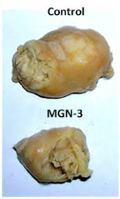
MGN-3/Biobran is a denatured hemicellulose obtained by reacting rice bran hemicellulose with multiple carbohydrate hydrolyzing enzymes from Shiitake mushrooms. Over the last 24 years, our fundamental research objective has been to study the biotherapeutic activity of MGN-3 as a treatment for cancer based on its ability to activate the immune system. This objective has been pursued in vitro, and in animal and human studies. This review is focused on the immunomodulatory effects of MGN-3 and on its potential as an anticancer agent. In vitro studies showed that culturing different human and murine cancer cell lines with MGN-3 resulted in a reduction of the survival rate of cancer cells. In vivo studies have also shown that MGN-3 induces tumor regression in several models of animal bearing tumor, including gastric cancer, neuroblastoma, and Ehrlich carcinoma. In addition, the anti-cancer activity of MGN-3 has been shown in human clinical trials and in several case reports on patients with Hepatocellular Carcinoma (HCC) and progressive and partially metastasized cancer. Patients that were treated with MGN-3 in addition to Conventional Therapy (CT), as compared with CT alone, showed: 1) less recurrence of cancer, 2) higher survival rate and 3) improved Quality of Life (QOL) as characterized by improvements in physical activity, appetite, sleep, and digestion, and a decrease in pain and anxiety.
This review summarizes the preclinical and clinical research on MGN-3/ Biobran since it was first patented in 1992. Various animal studies and human clinical trials including different types of malignancies have demonstrated that MGN-3 is a potent Biological Response Modifier (BRM). MGN-3 enhances the cytotoxic reactivity of immune cells with anti-cancer activity such as NK and CD8+ T cells via increasing cell granularity, stimulates the production of interferons, IL-2 and IL-12, and functions as a natural adjuvant for Dendritic Cells (DC). Therefore, MGN-3 may be used in DC-based vaccine strategies against infections and cancer. Importantly, MGN-3 is a unique BRM because it is a safe non-toxic agent and does not exhibit hyporesponsiveness. MGN- 3 has the potential to be a novel and promising immune modulatory adjuvant that could complement the existing immunotherapeutic modalities for cancer patients.
Despite the last decade of advances in treatment options, cancer remains the second leading cause of death in the United States. Unfortunately the outcome of standard cancer treatments is often poor due to the emergence of Multidrug Resistance (MDR) during the course of treatment. MDR cells are a significant factor in the failure of chemotherapeutics as evidenced by high relapse rates for the majority of patients. Therefore, to increase cancer survival and improve symptom control, there is a strong need for new and better approaches to cancer treatment. Today, the National Cancer Institute (NCI) has acknowledged the importance of immune therapy for the treatment of cancer. NCI, other health organizations, and professionals in the field of oncology are currently working to harness the immune system to fight cancer and to expand immunotherapy in combination with other types of cancer treatment, such as targeted therapy, chemotherapy, and radiation therapy.

Tuesday, 2 January 2018


The development in the field of bio sensing of biochemical molecules has been rapid during the recent years. Among many sensing technologies, Silicon Nanowire (SiNW)-based Field-Effect Transistors (FETs) have been shown to be as one of the most promising building blocks for the next generation electrical circuits in recognizing a wide range of biological and chemical targets. They have been successfully used in the detection of, for example, DNA, pH, protein, glucose, virus, and vapor. Despite the significant developments in the area, it seems, however, that the underlying detection mechanism and dynamics of the SiNW FETs are not well defined, and further studies are required. Due to the large surfaceto- volume ratio, one-dimensional nanostructures are considered as one of the best candidates for ultra-sensitive sensors. SiNW FETs have been experimentally demonstrated for direct, label-free, high sensitive, highly selective, and real-time detection of biological and chemical targets at very low concentrations. The most typical configuration of this device uses a nanowire as the essential building block bonding two ends of the nanowire to a solid substrate to create a SiNW FET.
Nanowire (NW) has attracted wide attention, and analyses that focus on various aspects of the device operation have accumulated. We have offered an experimental and analytical method to observe the poly silicon NW MOSFET threshold voltage based on a simple ballistic MOSFET modeling. This paper is intended to incorporate scattering effects into the ballistic modeling and to provide a compact model of the quasi-ballistic Si NW MOSFET. Silicon NW MOSFETs attract wide attention as a promising Nano device for future highdensity LSI application. For development of the device including the circuit application, a handy tool that affords accurate prediction of device characteristics is indispensable.
The device samples were manufactured on standard 6-in. p-type wafers. A proposed hybrid sensor/memory/CMOS poly-Si nanowire structure is illustrated in (Figure 1). The bottom-gate poly-Si nanowire formation can be inserted specifically after metallization of the back-end process (BEOL). At the beginning, buried oxide was deposited on a substrate surface as the gate dielectric of nanowire FETs. A 50-nm polysilicon layer was then deposited using the CVD process. Subsequently, the poly-Si wire was patterned by the standard I-line stepper of the CMOS semiconducting process. By using reactive plasma etching for photoresist trimming followed by silicon etching, the NW dimension was scaled to a level of approximately 100nm.

TB Treatment Success Rate in Ethiopia: Key Findings & Challenges

Tuberculosis (TB) remains a major global health issue , infecting one-third of the world's population . Despite efforts, Ethiopia's...